Section 2 | Respiratory Diseases in Horses: Rhodococcus equi
Industry
Page 08 /
Identifying Clinical Signs of Pneumonia in Foals
The focus of this FAAST Review is to discuss:
- How Rhodococcus equi pathogens are spread
- The different treatment options available
- Best practices to prevent pneumonia in foals from occurring
Infection with Rhodococcus equi is a leading cause of disease and death in foals. This FAAST Review is not intended to be provide exhaustive coverage of pneumonia-causing bacteria, but focuses on more common conditions, and ones where inappropriate antimicrobial use has lead to the development of resistance.
Signs of Rhodococcus equi
Rhodococcus equi most commonly presents as a case of bronchopneumonia (inflammation of the lungs) with abscess formation and infection of lymph nodes (a critical part of the body’s infection-fighting system).
Early Signs and Detection
Early clinical signs could include a slight cough, increase in breathing rate and a mild fever (>38.5°C or 101.3°F), however, these signs can be difficult to identify. Ultrasound is commonly used to screen the lungs for early detection. The veterinarian will look for the presence of lung consolidation (areas of the lung that have decreased capacity to take in air often due to fluid or pus accumulation) or abscesses in foals that do not show clinical signs1.
Some foals with changes identified on ultrasound may spontaneously recover without treatment. Work with your veterinarian to develop a protocol that works for you based on your farm history, geographic region, and age when foals are examined.
)
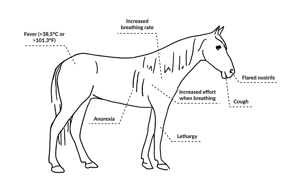
Clinical Signs
Clinical signs often present in foals that are less than 16 weeks of age.
Clinical signs include1:
- Fever (>38.5°C or >101.3°F)
- Lethargy
- Cough
- Anorexia
- Increased breathing rate
- Flared nostrils
- Increased effort when breathing
There are also some other manifestations of Rhodococcus equi infections, including:
- Diarrhea
- Multiple swollen joints
- Lesions on the foal’s eye
- Abscessed lymph nodes in the lungs and/or abdomen
)
Diagnosis of Disease
Your veterinarian may be suspicious of a Rhodococcus equi infection based on the results of the physical and/or ultrasound examination but confirming the diagnosis, requires the use of specialized tests (bacterial culture or a PCR from a tracheobronchial aspirate, where bacteria are identified under the microscope). Obtaining a sample for bacterial culture will also allow the determine the sensitivity of the bacteria to certain antimicrobials. These procedures are invasive so most veterinarians make the diagnosis based on ultrasound examination of the chest.
This should be completed when a foal has one or more of the following2:
- Clinical signs of lower respiratory disease
- Evidence of airway inflammation
- Presence of abscesses in the chest identified using an X-ray or ultrasound
These tests and indicators of disease require specialized training. Work with your veterinarian to develop a diagnostic plan for your foal if you suspect Rhodococcus equi is present
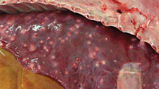
Abscesses in the lung of a foal affected by Rhodococcus equi
Source: Oklahoma Farm and Ranch
References:
- Giguere, S., N.D. Cohen, M Keith Chaffin, S.A. Hines, M.K. Hondalus, J.F. Prescott, and N.M. Slovis. 2011. Rhodococcus equi: Clinical manifestations, virulence, and immunity. J Vet Intern Med. 25.
- Giguere, S., N.D. Cohen, M Keith Chaffin, N.M. Slovis, M.K. Hondalus, S.A. Hines, and J.F. Prescott. 2011. Diagnosis, treatment, control, and prevention of infections caused by Rhodococcus equi in foals. J Vet Intern Med. 25:1209-1220.