What is American Foulbrood?
Industry
Page 04 /
Identifying Hives With American Foulbrood
Clinical Findings in American Foulbrood
There are numerous symptoms that suggest American Foulbrood (AFB) may be present when inspecting a hive. However, it can be difficult to determine AFB infection through these signs alone, as many other diseases can cause similar signs and symptoms.
As a result, it is important and required to report to the Provincial Apiarist / Ontario Apiary Program to arrange an inspection, as AFB is a reportable disease under the Ontario Bees Act. Beekeepers may also (in addition) wish to perform further testing in collaboration with their veterinarian to confirm the presence of AFB in their colony.
Source: Paul Kozak, OMAFRA
The findings most commonly found in hives affected with AFB include1,2:
Brood pattern (as seen in the figure below):
- The first sign observed with AFB is a spotty pattern of brood cells. Typically, there are few cells without larvae. This occurs due to the removal of infected brood by adult bees. Although, this can also occur:
- Due to other diseases such as European Foulbrood and varroa mites
- As a result of a poor queen
- Due to environmental factors, such as chilling and poor nutrition
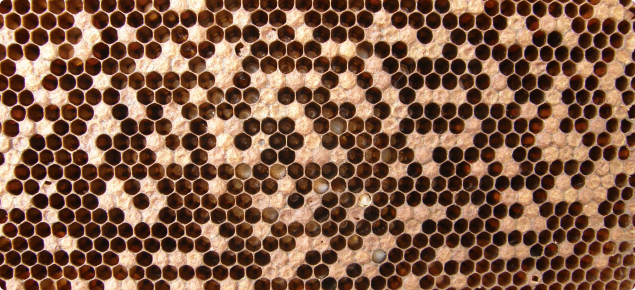
Figure: Brood pattern seen with AFB
Source: Bee Aware: American Foulbrood
Foul odour:
- A strong odour may be detectable due to the dead and decaying brood
Sunken caps:
- Larvae that are infected with AFB die after the caps are closed, which will cause the cap to shrink down and appear sunken
Perforated caps (as seen in figure below):
- AFB affected cells will develop dark, greasy, and sunken caps; they may become perforated by adult bees attempting to remove them. Brood disease should be considered when perforations are found around the edges of the cap. However, this is not always a sign of AFB, as holes in capping can occur in a healthy hive during the capping process, when bees are emerging from the cells, and due to hygienic behaviour
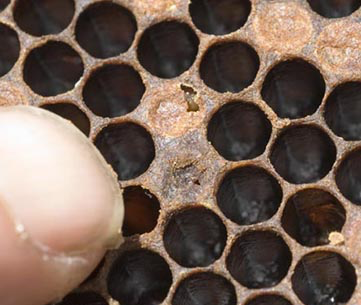
Figure: Perforated caps seen in hives infected with AFB
Source: Bee Aware: American Foulbrood
Larval scale (as seen in the figure below):
- Scale, consisting of dried, decomposed remains of larva or pupa, adheres tightly to the bottom of the cell walls making it difficult to be removed by bees
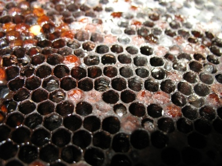
Figure: Dry scale
Source: OMAFRA
Pupal tongues (as seen in figure below):
- Dead pupae will lie flat at the bottom of the cells and the remnants of their tongues will extend upwards. While not a commonly seen feature, it is very indicative of AFB
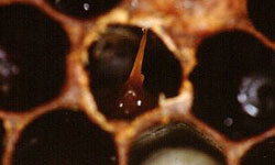
Figure: Presence of pupal tongues due to AFB
Source: Bee Aware: American Foulbrood
Colour change:
- The infected brood changes in colour from white to dark brown
Remember: As many of these signs are present with diseases other than AFB, it is important and required to report to the Provincial Apiarist to arrange an inspection if you suspect that AFB is affecting your hive. You may wish to perform further testing in collaboration with your veterinarian to confirm the presence of AFB
Diagnosis of American Foulbrood
It is important to detect AFB early and eradicate it as soon as possible. Beekeepers should inspect their brood nests regularly throughout the season (at least four times a year), and before the application of any treatments, which may mask its symptoms.
Although some of the clinical symptoms described previously can be used to diagnose AFB in the field, not all the clinical signs are specific to AFB. It is important and required to report to the Provincial Apiarist / Ontario Apiary Program to arrange an inspection if you suspect that AFB is present, as it is a reportable disease under the Ontario Bees Act. Ontario Beekeepers may also (in addition) wish to perform further testing in collaboration with your veterinarian to confirm the presence of AFB in your colony.
Matchstick/rope test:
- To complete this test, take a matchstick or toothpick and put it into a cell that has a discoloured cell cap and slowly pull it out. If the decaying product in the cell pulls or ropes out and is 2 cm or more in length with a mucus-like consistency then it suggests the presence of AFB. A negative test (less than 2 cm of decaying product roping out) does not rule out AFB and more invasive testing will need to be completed. Note that this test does not work with dry AFB scale
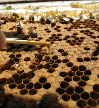
Source: OMAFRA
Enzyme linked immunosorbent assay (ELISA) test:
- Working with your veterinarian, a field ELISA test may also be completed. Your vet will help you to interpret the results and be able to provide recommendations on how best to proceed
Laboratory testing:
- Working with your veterinarian, samples of the brood comb can be sent into a laboratory for more intensive testing to confirm the presence of AFB under the microscope or through bacterial culture
- The Animal Health Laboratory at the University of Guelph requires a 10 cm x 10 cm piece of brood comb, 40 mL of honey, 50 dead bees, and wax scrapings for diagnosis through bacterial culture
References
- De Graaf, D.C., A.M. Alippi, M. Brown, J.D. Evans, M. Feldlaufer, A. Gregorc, M. Hornitzky, S.F. Pernal, D.M.T. Schuch, D. Titera, V. Tomkies, and W. Ritter. 2006. Diagnosis of American Foulbrood in honey bees: A synthesis and proposed analytical protocols. Letters in Applied Microbiology. 43:583-590.
- Genersch, E. 2010. American Foulbrood in honeybees and its causative agent, Paenibacillus larvae. J Invertebrate Pathology. 103:10-19.
- USDA. Role of veterinarians in honey bee health.